Electron-Microprobe Lab
Contact:
Dirk Müller
Dirk.Mueller@min.uni-muenchen.de
Telefon: +49 (0)89 2180-4276
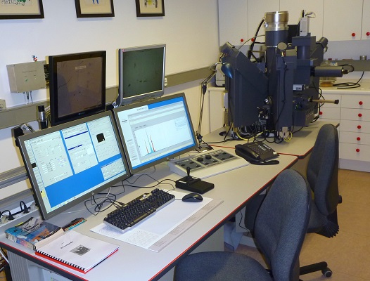
The Cameca SX100 electron probe microanalyser (EPMA) used at the Department of Earth & Environmental Sciences is a fully automated instrument employing five wavelength dispersive spectrometers for the detection and nondestructive analysis of almost all elements from boron down in the periodic table. Quantitative analysis may be carried out on the bulk matrix and micro-constituents, such as inclusions and grain boundaries in metals and intermetallic phases, geological materials, coal, ceramics, glasses and building materials. X-ray and electron images may be acquired across constituents of interest at high lateral resolution. Special procedures were developed to meet the requirements of a reliable analysis for different materials:
A common defect in single crystal growth from solution is the formation of mother-liquid inclusions. In order to understand their evolution and improve the experimental procedure it is crucial to know their chemical and structural composition. Their fine-grained (< 5 µm) internal structure demands special requirements for EPMA measurements, realized by low-voltage (5 keV) elemental mapping applying a step size of 0.138 µm for each pixel (Müller et al. 2014).
Alkali-ion migration out of the excitation volume has been a well-known challenge in electron microanalysis for the last decades. For the analysis of alkali-rich silicate glasses it is a necessary requirement to prevent this migration process. This could be successfully done by using an optimized operating protocol, applying beam current densities lower than 0.1 nA/µm² (Scherrer 2012).
The groundmass of steam cured building materials consists of calciumsilicate-hydrates as tobermorite and semicrystalline CSH(I). Their grain sizes are as low as 5 µm and they exhibit thermal instability under the electron beam. Knowledge of their composition is significant in order to determine the thermodynamic and kinetic properties of tobermorite-forming reactions. High resolution can be achieved by scanning the beam over a small area of 60x60 µm by concomitant reduction of heating up of the sample. By applying a special in-house developed beam-scanning method not only the composition, but also the amount of individual calcium-silicate-hydrates can be determined (Fehr & Zürn 2000).
selected publications:
- Müller D, Schwerin J, Gille P, Fehr KT (2014): High-Resolution EPMA X-ray Images of Mother Liquid Inclusions in a Pd2Ga Single Crystal, IOP Conference Series: Materials Science and Engineering, 55, 012013. http://dx.doi.org/10.1088/1757-899X/55/1/012013
- Scherrer M (2012): Characterisation of synthetic alkali-rich silicate melts by EPMA and viscometry, MSc thesis, LMU München.
- Fehr KT, Zürn SG (2000): Determination of CSH-phases by beam-scanning electron microprobe techniques. In: Rammelmair D, Mederer J, Oberthür Th, Heimann RB, Pentinghaus H (eds.): Applied Mineralogy in Research, Economy, Technology, Ecology and Culture, A.A. Balkema, Rotterdam, 761-764.